Insufficient oxygen supply to the heart muscle, often caused by conditions like coronary artery disease or respiratory disorders, can lead to impaired cardiac function and decreased cardiac output. Conditions that affect the structure and function of the heart muscle, such as cardiomyopathy or myocarditis, can impair the heart’s ability to pump effectively, resulting in decreased cardiac output. Any disturbance in the heart’s electrical system can disrupt the coordinated contraction and relaxation of the cardiac muscle, leading to decreased cardiac output. Alteration in heart rate, rhythm, and conduction.Additionally, here are some related factors that may be related to decrease in cardiac output: Several cardiovascular disease such as myocardial infarction, heart failure, dysrhythmias, and other problems in fluid volume, can cause decrease in cardiac output Causes of decrease in cardiac outputĬonditions like myocardial infarction, hypertension, valvular heart disease, congenital heart disease, cardiomyopathy, heart failure, pulmonary disease, arrhythmias, drug effects, fluid overload, decreased fluid volume, and electrolyte imbalance is common causes of decreased cardiac output.

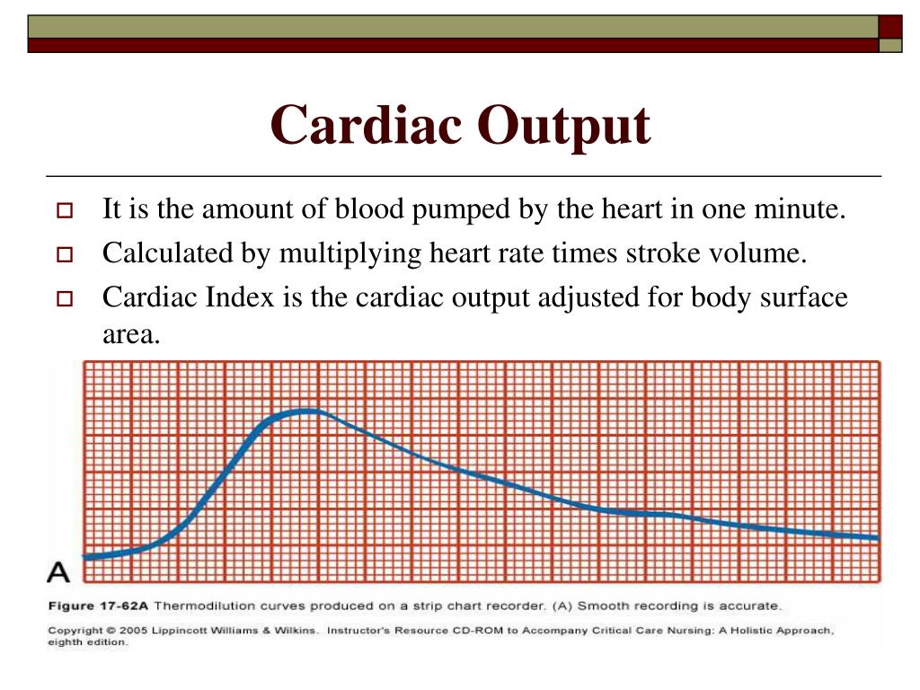
The cardiac output is calculated using an algorithm based on the pulse waveform.Ī decrease in cardiac output occurs when the blood pumped by the heart does not meet the metabolic demands of the body. Is a technique that involves using a specialized device to measure the shape and strength of the arterial pulse wave. Using a specialized thermistor-tipped catheter, the temperature change can be used to calculate the cardiac output. This method involves injecting a small amount of cold saline into an artery via a PAC and measuring the temperature change in the blood as it flows through the body. The technique is based on Fick’s principle which states that the rate of oxygen consumption is equal to the product of the cardiac output and the difference in oxygen content between the arterial and venous blood. This method involves measuring the oxygen consumption of the body and the amount of oxygen being delivered to the body. This shift is used to determine flow velocity and flow volume which are used to calculate for cardiac output using a formula. The velocity of the blood through the heart causes a Doppler shift in the frequency of returning ultrasound waves. This method uses ultrasound and the doppler effect to measure cardiac output.

There are several methods that can be used to calculate and measure cardiac output, including the Doppler ultrasound, Fick Method, the thermodilution method, and arterial pulse contour analysis method. For example, elite athletes have a cardiac output of more than 35L/min during exercise. However, it is important to note that normal range can vary depending on various factors such as age, size, and activity level. The normal cardiac output of a healthy adult is generally considered to be between 5-6 liters per minute (L/min) at rest (King and Lowery, 2022). The cardiac output is usually expressed in liters/minute (L/min). It is the product of the heart rate, which is the number of beats per minute, and the stroke volume, which is the amount pumped per beat (Cardiac output = heart rate x stroke volume). Cardiac output is the amount of blood pumped by the heart per minute.


 0 kommentar(er)
